This book helps the reader to interpret the reproductive status of the cow using an outstanding selection of photographs with an eminently practical focus. The carefully chosen images let the reader visualize what they are touching during an examination, and describe the more basic procedures, such as rectal palpation or artificial insemination. It also highlights the importance of using oestrus synchronization protocols in routine reproductive practice.
The Oestrous Cycle of the Cow A photographic atlas UPDATED EDITION
This book helps the reader to interpret the reproductive status of the cow using an outstanding selection of photographs with an eminently practical focus. The carefully chosen images let the reader visualize what they are touching during an examination, and describe the more basic procedures, such as rectal palpation or artificial insemination. It also highlights the importance of using oestrus synchronization protocols in routine reproductive practice.
KEY POINTS:
➜ Includes many visual materials that are of great educational value.
➜ Relates reproductive success to successful production, and describes basic procedures such as rectal palpation and artificial insemination.
➜ Helps the reader create a decision tree that will help them diagnose the reproductive stage of the cow being examined.
Author
MANUEL FERNÁNDEZ SÁNCHEZ
Veterinary sciences graduate of the University of Zaragoza, Spain. He currently coordinates nutrition projects, and specialises in analysing data generated on farms through various online initiatives. He is the webmaster of consuvet.com and the creator of www.cowsulting.com, www.recetaveterinaria.com, and www.cowculations.com
Table of contents
1. The reproductive tract of the cow Morphology, structure and function Position Reproductive organs of the cow Anatomy of the ovary Anatomy of the oviduct Anatomy of the uterus. Horns and body Anatomy of the uterus. Cervix Function of the ovary Hypothalamic-pituitary axis Relationship between production and reproduction Examination of the reproductive tract by rectal palpation Artificial insemination
2. Oogenesis, follicular development and dynamics Oogenesis Follicular development Follicular dynamics Synchronisation protocols
3. Recognising structures Guide to identify ovarian structures General evaluation of the cycle Recognising ovarian structures
4. The puerperium
5. Pregnancy diagnosis by palpation
6. Ovarian cyst Follicular cyst Luteal cyst Persistent follicle Post-partum anoestrus Metritis Mummified foetuses Embryonic reabsorption Other processes
Data sheet
Specific References
The volume has been updated and enriched by the Gastroenterology academics to offer a user-friendly tool, always in line with the most recent research and pedagogical methodologies.
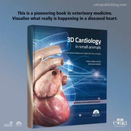
This is a pioneering book in veterinary medicine. The reader will be able to visualize what really is happening in a diseased heart. Through three-dimensional representation and augmented reality; every disorder can be seen, almost touching the 3D model above the book. The pathologies' changes in blood flow can be visualized, bringing the reader even closer to the pathophysiological reality. The text addresses the major points of pathophysiology and diagnostic keys for the most common diseases, and the illustrations and pictures are a bridge between the 3D models and the text. Thus, the book succeeds in joining diagnosis, particularly a test such as echocardiography, to understand the pathophysiology.
Professional responsibility, “good practice and malpractice” in obstetrics is one of the major areas of medical-legal litigation.
This visual atlas takes a trip through the digestive system, describing general concepts of its anatomy and physiology. It covers the diagnostic and therapeutic approach to common disorders of the gastrointestinal tract, hepatobiliary tract, and pancreas. Enriched with simple yet comprehensive illustrations, it serves as a tool to facilitate communication between veterinarians and pet owners.
THE BOOK IS AVAILABLE FOR SHIPMENT STARTING ON 15 NOVEMBER
Microsurgical Endodontics, a term coined by the author himself, is a branch of Endodontics that allows to save dental elements otherwise lost and intended to be replaced with implants. The study of it means adding precious information to your knowledge in endodontics. There are many indications of intervention, always linked to the failure of a previous treatment and may include solutions that, once put into practice, can resolve clinically complicated situations in a single session. The text presents a complete examination of the diagnostic approaches, of the clinical procedures, of the necessary tools and of the possible complications that Microsurgical Endodontics involves.
This book helps the reader to interpret the reproductive status of the cow using an outstanding selection of photographs with an eminently practical focus. The carefully chosen images let the reader visualize what they are touching during an examination, and describe the more basic procedures, such as rectal palpation or artificial insemination. It also highlights the importance of using oestrus synchronization protocols in routine reproductive practice.