
In recent years, musculoskeletal ultrasound (MSUS) has achieved an important role not only in clinical applications (for diagnosis and management of various musculoskeletal disorders), but also in research, due to its several advantages (being convenient, inexpensive, non-invasive, repeatable, providing high-resolution dynamic and comparative imaging, and not requiring any exposure to radiation).
In recent years, musculoskeletal ultrasound (MSUS) has achieved an important role not only in clinical applications (for diagnosis and management of various musculoskeletal disorders), but also in research, due to its several advantages (being convenient, inexpensive, non-invasive, repeatable, providing high-resolution dynamic and comparative imaging, and not requiring any exposure to radiation). As such, MSUS has increasingly become a valuable tool in the daily clinical practice of Physical and Rehabilitation Medicine (PRM) physicians and, where used in a rehabilitation setting, it can significantly contribute to the diagnostic and therapeutic algorithm of rehabilitation patients. For these reasons, the MSUS probe can be now thought of as synonymous with the physician’s stethoscope. Additionally, scanning is quite comfortable, and sometimes even more reassuring for the patient than the physician: “seeing is believing”. In this book, aside from drawing attention to the growing issues in the agenda of PRM physicians using MSUS, the authors also discuss basic technical features of the technique and focus on both diagnostic and interventional utility of MSUS in different health conditions involving muscles, tendons, ligaments, nerves, and joint lesions.
Authors
Levent Özçakar and Martine De Muynck
Levent Özçakar, Professor at Hacettepe University Medical School, Department of Physical and Rehabilitation Medicine, Ankara, Turkey. His main areas of interest are musculoskeletal ultrasonography and thoracic outlet syndrome. He is the founder and President of TURK- MUSCULUS (Turkish Musculoskeletal Ultrasound Study Group), EURO-MUSCULUS (European Musculoskeletal Ultrasound Study Group), WORLD-MUSCULUS (World Musculoskeletal Ultrasound Study Group), TURK-MUS (Turkish Multidisciplinary Ultrasound Society), USPRM (Ultrasound Study Group of ISPRM), and ESPRM (European Society) Special Interest Committee on Musculoskeletal Ultrasound. He is also the national delegate for EULAR Standing Committee on Musculoskeletal Imaging. He has authored over 370 publications in various international journals indexed by SCI/SCI-Exp. He is also Editor, member of the Editorial Board and/or reviewer for many international journals indexed by SCI/SCI-Exp.
Martine De Muynck, Head of the Department of Physical Medicine and Rehabilitation, Gent University Medical School and Professor at the Gent University Medical School, Belgium. Her main areas of interest are musculoskeletal ultrasound and electrophysiology of the pelvic diaphragm. She has actively participated in the Royal Belgian Society of Physical and Rehabilitation Medicine for many years, as a secretary and as the first female President (2005). He has organized several national and international seminars on ultrasound and gave lectures in national and international congresses and meetings. She is a co-founder of the EURO-MUSCULUS (European Musculoskeletal Ultrasound Study Group).
Data sheet
Specific References
Comprehensive, specialised and practical atlas useful for anyone who is starting out or who wants to delve deeper in the differential aspects of the cat as a surgical patient. It covers the most common surgical interventions in the feline species which are duly explained through photographs and detailed illustrations. The authors have many years of experience in feline medicine and surgery, and their goal is to impart their knowledge in the most practical way possible.
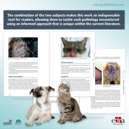
Servet presents this innovative work written by authors specialised in the field. Uniquely, this book provides a joint perspective on the subjects of Dermatology and Immunology. The combination of the two subjects makes this work an indispensable tool for readers, allowing them to tackle each pathology encountered using an informed approach that is unique within the current literature.
The second edition of this educational atlas on cats expands on its initial goal of helping veterinary surgeons communicate with owners by adding to the content of the first edition with 16 new sheets. Needless to say that the previous topics have been revised and updated with the latest developments in each area.
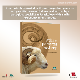
Atlas entirely dedicated to the most important parasites and parasitic diseases of sheep, and written by a prestigious specialist in Parasitology with a wide experience in this species. More than 300 high-quality images have been included to show, among other things, the main parasites (protozoa, helminths and arthropods) infecting sheep, providing identification features to clearly distinguish between species, as well as the principal clinical signs derived from each infection. Each chapter has been dedicated to a specific anatomic system of sheep.
In recent years, musculoskeletal ultrasound (MSUS) has achieved an important role not only in clinical applications (for diagnosis and management of various musculoskeletal disorders), but also in research, due to its several advantages (being convenient, inexpensive, non-invasive, repeatable, providing high-resolution dynamic and comparative imaging, and not requiring any exposure to radiation).