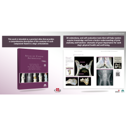

In this practical atlas, the description of the anatomy of the dog’s joints provided in the previous edition has been completed and complemented with new tools, 3D animations, and self-evaluation tests that will help readers acquire knowledge and form a better understanding of joint anatomy and function – elements of great importance for each dog’s physical health and well-being.
This work is intended as a practical atlas that provides a comprehensive description of the anatomy of each component found in a dog’s articulations. However, in this updated edition of Atlas of Canine Arthrology, the information provided in the previous version has been enhanced and complemented with new tools, 3D animations, and self-evaluation tests that will help readers acquire knowledge and form a better understanding of joint anatomy and function– elements of great importance for each dog’s physical health and well-being.
KEY FEATURES:
➜ Detailed description of each joint accompanied by high-quality graphical material.
➜ High quality illustrations.
➜ 3D animations that allow the reader to visualise the joints in all possible positions.
➜ Self-assessment tests that allow readers to consolidate their knowledge.
Authors:
JESÚS LABORDA VAL
Professor at the Department of Animal Anatomy, Embryology, and Genetics (University of Zaragoza’s Faculty of Veterinary Medicine) since 1989.
JULIO GIL GARCÍA
Lecturer at the Department of Animal Anatomy, Embryology, and Genetics (University of Zaragoza’s Faculty of Veterinary Medicine).
MIGUEL GIMENO DOMÍNGUEZ
Lecturer at the Department of Animal Anatomy, Embryology, and Genetics (University of Zaragoza’s Faculty of Veterinary Medicine).
Table of Contents
1. General aspects
Angles and joints
Characteristics of articular cartilage
Ossification of the thoracic limb
Ossification of the pelvic limb
Characteristics of the synovial joint
2. Head and axial region
Joints of the head
Joints of the vertebral column
Joints between vertebrae and ribs
Joints of the sternum
Joints of the thoracic limb
Joints of the pelvic limb
Self-assessment
3. Thoracic limb
Joints
Self-Assessment
4. Pelvic limb
Joints
Self-assessment
Data sheet
Specific References
Who said that nutrition is boring? Not in this book! Here we have transformed nutrition into a series of graphic pages, with simple and concise text and several diagrams, drawings and images, by which the veterinarian can see all of the fundamental aspects of nutrition in dogs and cats. From digestive physiology and dietary behavior, the evaluation of body condition and morphometric measurements, to the characterization of the immediate principles and the description of the energy and nutrient requirements, this atlas is a succession of pages with an immense richness in information and visuals that will enable the reader to enjoyably deepen their knowledge about pet nutrition.
The centrality of nutrition in the state of health of pets has emerged only in the recent years, both to prolong their life expectancy and to prevent the onset of serious diseases such as obesity, diabetes mellitus or liver lipidosis. The goal of this book is that each veterinarian can clearly answer the questions that are most frequently asked by the owners: Which type of food to choose? How to navigate between the different products on the market? The text also provides real recipes to be proposed in synergy with the nutritional handbooks of various food manufacturers, to consciously choose and be able to compare the nutritional characteristics of different products on the market.
The aim of this book is to focus on problems unique to the feline alimentary tract in comparison to dogs and to discuss them in detail, but also to highlight areas where knowledge is lacking or can only be derived from comparison with other companion animal species or humans. Instead of being a comprehensive work of “all things GI” in the cat, this book aims to shine a light on topics that are novel, such as the microbiome or probiotics, and might not have been covered by other standard textbooks. This book focuses on “a medic’s perspective” on feline alimentary tract health, which starts with considering differential diagnoses in a structured way based on the most common clinical signs.
After the introduction in the 1980s of minimally invasive surgery in human medicine, laparoscopic and thoracoscopic surgery started to take hold also in veterinary medicine. Thanks to its reduced invasiveness and its fast post-operative recovery, minimally invasive surgery started to expand so quickly that nowadays it has become the preferred technique for many surgical procedures. Laparoscopy and Thoracoscopy in the dog and cat is a comprehensive book that describe all the minimally invasive surgical procedures.
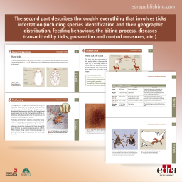
Very visual handbook aimed at veterinary professionals and dealing with everything related to fleas and ticks, focusing particularly on clinical issues in dogs and cats. The first part is entirely dedicated to fleas (including their identification, life cycle, clinical signs derived from flea infestation, flea allergy dermatitis (FAD), and prevention and control measures, among others). The second part describes thoroughly everything that involves ticks infestation (including species identification and their geographic distribution, feeding behaviour, the biting process, diseases transmitted by ticks, prevention and control measures, etc.).
In this practical atlas, the description of the anatomy of the dog’s joints provided in the previous edition has been completed and complemented with new tools, 3D animations, and self-evaluation tests that will help readers acquire knowledge and form a better understanding of joint anatomy and function – elements of great importance for each dog’s physical health and well-being.