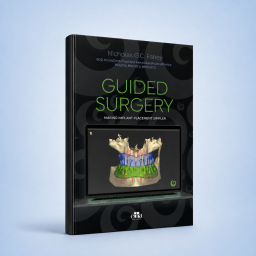
GUIDED SURGERY Making...
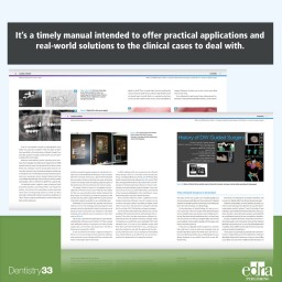
This is not the ordinary academic text to have in your library. It’s the result of the commitment of the Author, a practicing clinician who has focused his career on practical solutions to treat thousands of cases.
Active filters
This is not the ordinary academic text to have in your library. It’s the result of the commitment of the Author, a practicing clinician who has focused his career on practical solutions to treat thousands of cases.
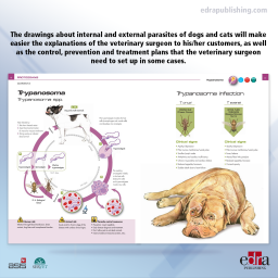
This illustrated work has the aim to assist the veterinary surgeon with his/her communication with the pet owner. Following the last book spirit dedicated to surgery, in this atlas, the drawings about internal and external parasites of dogs and cats will make easier the explanations of the veterinary surgeon to his/her customers, as well as the control, prevention, and treatment plans that the veterinary surgeon needs to set up in some cases. The clarity and accuracy of the drawings, designed to make it understandable and to reduce the time spent on the explanations to the pet owner, make this atlas another useful clinical tool. This book is aimed at veterinary surgeons, students, teachers, and professionals in this field.
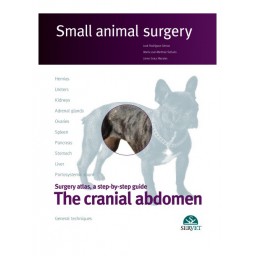
Servet presents the third volume of the publication “Small animal surgery”, which describes the main surgical procedures on the cranial abdomen of the dog, cat and ferret. This work, which has been acclaimed by professionals, stands out for the excellence of its images and the step-by-step description of each surgical technique using real clinical cases.
Diagnostic imaging is a key component in the care of companion animal patients. In recent decades the available imaging modalities have expanded from conventional radiography to include ultrasonography, computed tomography, and magnetic resonance imaging, and all are employed on a daily basis in both general and specialty veterinary practice.