
0
CART
0 item
Categories
-
Veterinary Books
-
Companion Animals
- Anaesthesia/Analgesia
- Anatomy
- Cardiology
- Cytology/Laboratorial
- Dermatology
- Diagnostic imaging
- Emergency
- Endocrinology
- Endocrinology/ Reproduction
- Equine
- Ethology
- Infectious diseases and immunology
- Nephrology/Urology
- Nutrition
- Oncology
- Ophthalmology
- Parasites
- Pet owner educational atlas
- Rehabilitation
- Senior care
- Surgery
- Surgery and traumatology/Orthopaedics
- Veterinary Dentistry
- Livestock
- Management
-
Companion Animals
- Dentistry Books
- Medicine Books
- CE Webinar
- CE Webinar
- French books
- E-books
Categories
-
Veterinary Books
-
Companion Animals
- Anaesthesia/Analgesia
- Anatomy
- Cardiology
- Cytology/Laboratorial
- Dermatology
- Diagnostic imaging
- Emergency
- Endocrinology
- Endocrinology/ Reproduction
- Equine
- Ethology
- Infectious diseases and immunology
- Nephrology/Urology
- Nutrition
- Oncology
- Ophthalmology
- Parasites
- Pet owner educational atlas
- Rehabilitation
- Senior care
- Surgery
- Surgery and traumatology/Orthopaedics
- Veterinary Dentistry
- Livestock
- Management
-
Companion Animals
- Dentistry Books
- Medicine Books
- CE Webinar
- CE Webinar
- French books
- E-books

Active filters

Practical Small Animal...
Price
CAD 127.00
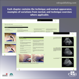
This book aims at being a quick visual guide to abdominal ultrasound in small animals. Each chapter contains the technique and normal appearance, examples of variations from normal, and technique exercises where applicable. The digital version includes scanning technique videos.
Immunosuppressive...
Price
CAD 120.75
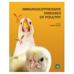
The goal of this work is to present the reader with the most common diseases that can produce immunosuppression in poultry, provide guidance for the diagnosis of immunosuppressive diseases, and examine the challenges that a diagnostician may face in confirming a diagnosis of immunosuppression. The book will feature an extensive collection of color photographs depicting gross and microscopic images.
Practical Small...
Price
CAD 101.85
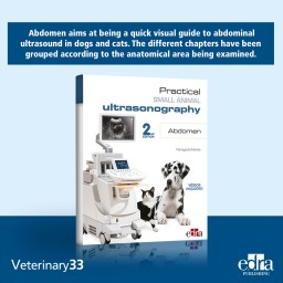
Practical small animal ultrasonography. Abdomen aims at being a quick visual guide to abdominal ultrasound in dogs and cats. The different chapters have been grouped according to the anatomical area being examined. Each chapter contains the technique and normal appearance, examples of variations from normal, and technique exercises where applicable. Quite a few images per chapter and videos with scanning techniques enrich this practical work. 2nd edition includes up-to-date content, a new section on portosystemic shunts and new video.

Atlas Of Radiographic...
Price
CAD 153.30
This second edition of the book enhances its original goal of aiding veterinary practitioners in radiographic interpretation and pathological diagnosis. It builds upon the first edition by introducing a chapter on radiology principles and a comprehensive atlas for radiographic positioning and anatomy. The edition includes 400 new images and has updated existing graphics to improve quality. Additionally, the book features new multimedia content accessible via QR codes in the text, allowing readers to view diagrams of normal radiographic anatomy, both labeled and unlabeled, for better understanding and reference.