


Atlas of Embryonic...
Price
CAD 51.45
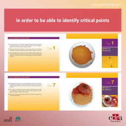
A visual atlas which describes –day by day– the hole process of embryonic development in order to be able to identify critical points in this process so a final and correct diagnosis can be established.