
0
CART
0 item
Categories
-
Veterinary Books
-
Companion Animals
- Anaesthesia/Analgesia
- Anatomy
- Cardiology
- Cytology/Laboratorial
- Dermatology
- Diagnostic imaging
- Emergency
- Endocrinology
- Endocrinology/ Reproduction
- Equine
- Ethology
- Infectious diseases and immunology
- Nephrology/Urology
- Nutrition
- Oncology
- Ophthalmology
- Parasites
- Pet owner educational atlas
- Rehabilitation
- Senior care
- Surgery
- Surgery and traumatology/Orthopaedics
- Veterinary Dentistry
- Livestock
- Management
-
Companion Animals
- Dentistry Books
- Medicine Books
- CE Webinar
- CE Webinar
- French books
- E-books
Categories
-
Veterinary Books
-
Companion Animals
- Anaesthesia/Analgesia
- Anatomy
- Cardiology
- Cytology/Laboratorial
- Dermatology
- Diagnostic imaging
- Emergency
- Endocrinology
- Endocrinology/ Reproduction
- Equine
- Ethology
- Infectious diseases and immunology
- Nephrology/Urology
- Nutrition
- Oncology
- Ophthalmology
- Parasites
- Pet owner educational atlas
- Rehabilitation
- Senior care
- Surgery
- Surgery and traumatology/Orthopaedics
- Veterinary Dentistry
- Livestock
- Management
-
Companion Animals
- Dentistry Books
- Medicine Books
- CE Webinar
- CE Webinar
- French books
- E-books


Active filters

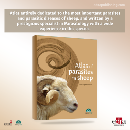

Atlas of Parasites in...
Price
CAD 116.55
Atlas entirely dedicated to the most important parasites and parasitic diseases of sheep, and written by a prestigious specialist in Parasitology with a wide experience in this species. More than 300 high-quality images have been included to show, among other things, the main parasites (protozoa, helminths and arthropods) infecting sheep, providing identification features to clearly distinguish between species, as well as the principal clinical signs derived from each infection. Each chapter has been dedicated to a specific anatomic system of sheep.
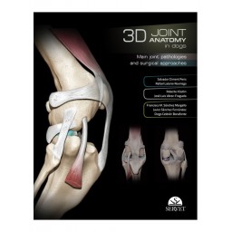
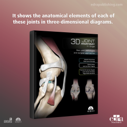
3D Joint Anatomy in...
Price
CAD 132.30
A visual guide with a strongly educational approach covering the main joints in the limbs of the dog. It shows the anatomical elements of each of these joints in three-dimensional diagrams. The views chosen for each case have been selected for a practical purpose, showing the position of the elements involved in the most commonly used surgical approaches. It also describes the key orthopaedic conditions affecting each joint and the most commonly used surgical approaches. It contains a large number of images and illustrations, and a selection of views presented in digital video format.

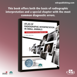
Atlas of Radiographic...
Price
CAD 120.75
This book, structured according to the normal anatomical areas in radiological diagnostic imaging: abdomen, neck, thorax, limbs, spine, and head, offers the key aspects of both radiographic interpretation and the diagnosis of diseases, as well as a special chapter with the most common diagnostic errors. Its more than 500 high-resolution images are accompanied by the necessary text to increase their descriptive value. In addition, the book is complemented by multimedia material, which can be accessed through QR codes located throughout the text. In this way, the reader can access different normal radiographic anatomy diagrams, both with and without the anatomical details identified. All these elements make this book a reference in the field of clinical radiology.


Atlas of Embryonic...
Price
CAD 51.45
A visual atlas which describes –day by day– the hole process of embryonic development in order to be able to identify critical points in this process so a final and correct diagnosis can be established.